Research

How microglia and macrophages shape the brain and body.

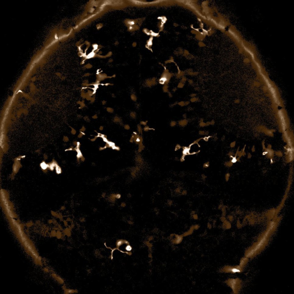
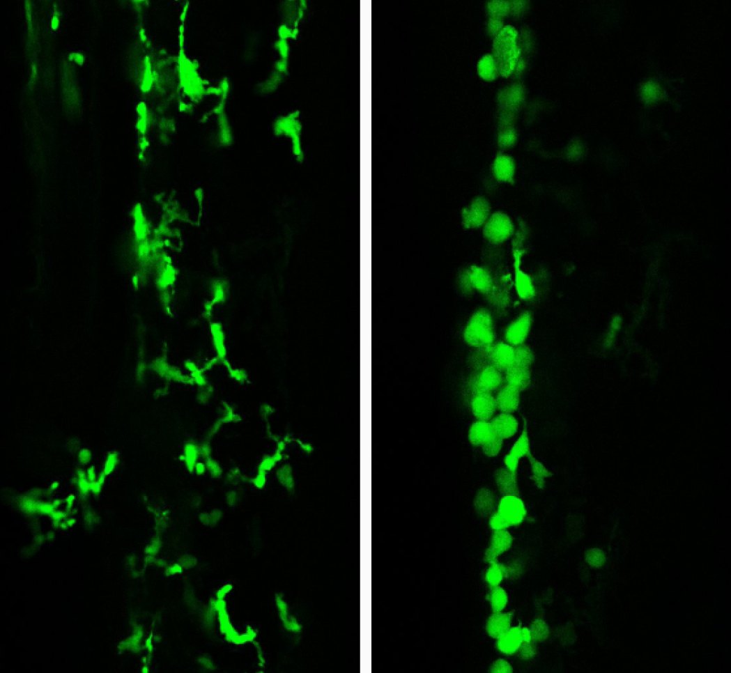
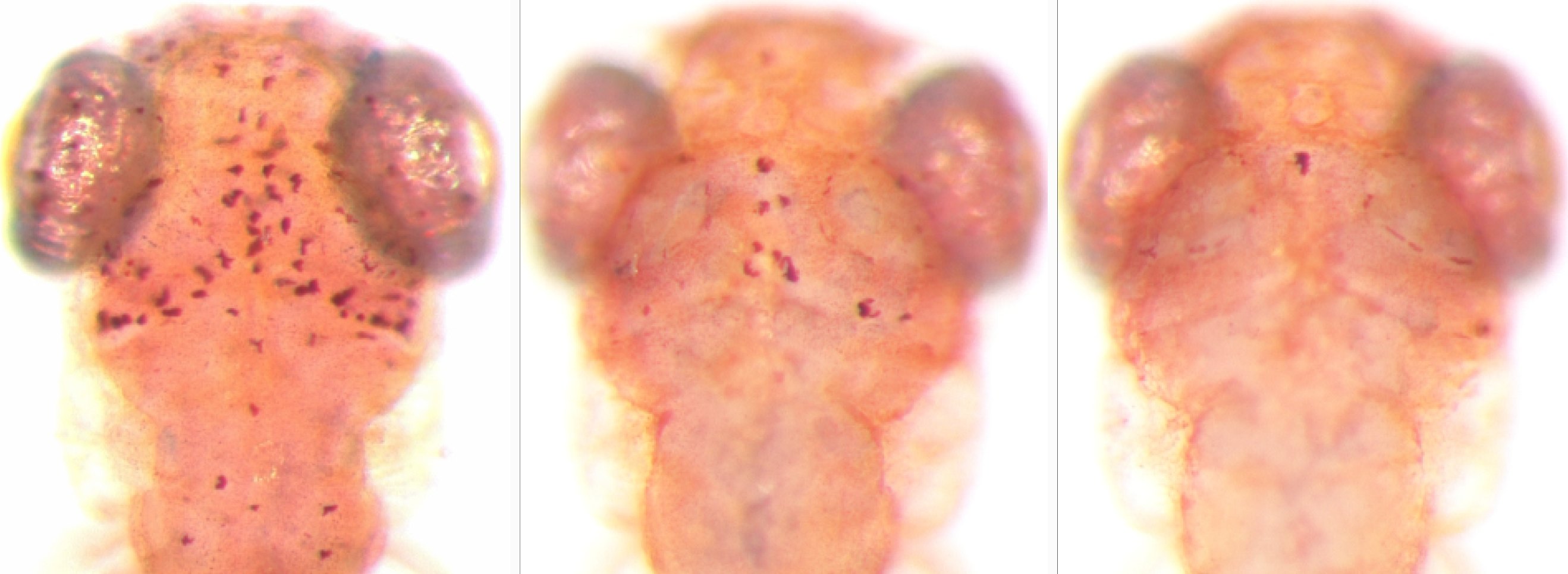
We combine live imaging, genetics, cell biology, and evolution in zebrafish and cavefish to understand the brain’s immune cells: how they work, how they evolved, and what goes wrong in disease.

Model systems

Why fish?

Microglia lose much of their identity outside the brain, so we study them where they patrol: inside the intact, living animal. Two small fish let us watch them at work. We then make cross-species comparisons, validating key observations from these teleosts in human cell lines to test whether the mechanisms we uncover are conserved.

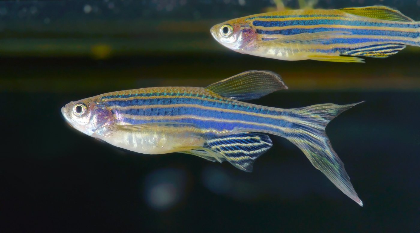
Two adult zebrafish, with blue and gold horizontal stripes, against a dark background

Zebrafish

Other than the fact that we think zebrafish are simply the best, they are ideal for studying microglia. Their embryos are transparent, so we can follow microglia and the developing neuroimmune system cell by cell with live, in vivo imaging. Each pair lays hundreds of eggs, which lets us run genetic screens of genes linked to neurodegenerative, neurodevelopmental, and neuropsychiatric disorders.

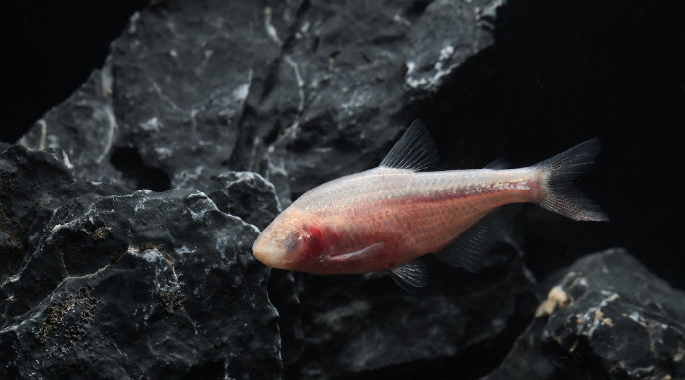
A pale pink, eyeless cavefish swimming past dark rocks

Mexican tetra

Astyanax mexicanus lives as eyed surface fish in rivers and as blind cavefish in caves. Many traits that differ between the surface and cave morphotypes converge on microglia and macrophages. We are using our expertise in zebrafish macrophage biology to understand how microglia and macrophages evolved in the Mexican tetra.

Disease

Microglia as contributors to neurological disease.

Microglia are the only resident immune cells of the brain parenchyma, and they are involved in nearly every neurological disorder. Yet research has long centered on neurons, leaving open how microglia shape, protect, and sometimes damage the brain. Because microglia are long-lived, self-renewing, and cannot be replaced by healthy cells from the blood, a change in their state early in life can permanently alter brain wiring.

The role of microglia is especially understudied in pediatric conditions. For example, in lysosomal storage disorders, rare neurodegenerative diseases of childhood, microglial activation precedes and predicts where neurons will be lost, and bone marrow transplants help some children, pointing to the immune system as a driver of disease.

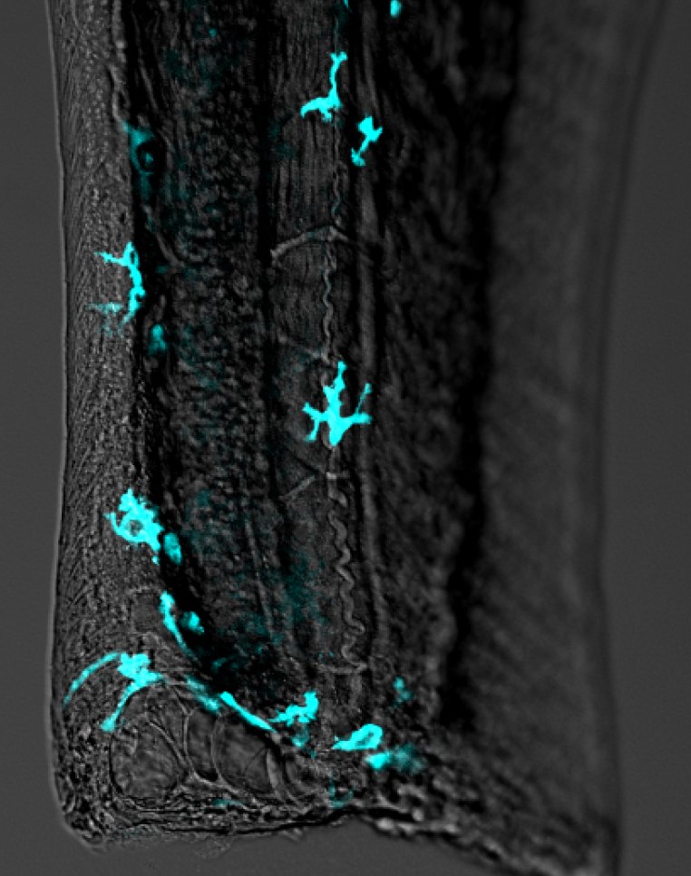
Zebrafish larvae develop rapidly in a dish, allowing us to uncover the earliest disease-associated changes in nervous system formation and function. The optical transparency of the embryos also enables tracking of individual cell types in the brain. Importantly, because zebrafish are ideal for rapid CRISPR screens, we can test many disease-associated genes at once, from pediatric lysosomal storage disorders and neurodevelopmental conditions to Alzheimer’s disease. We then follow the strongest candidates into how microglia impact disease onset and progression.

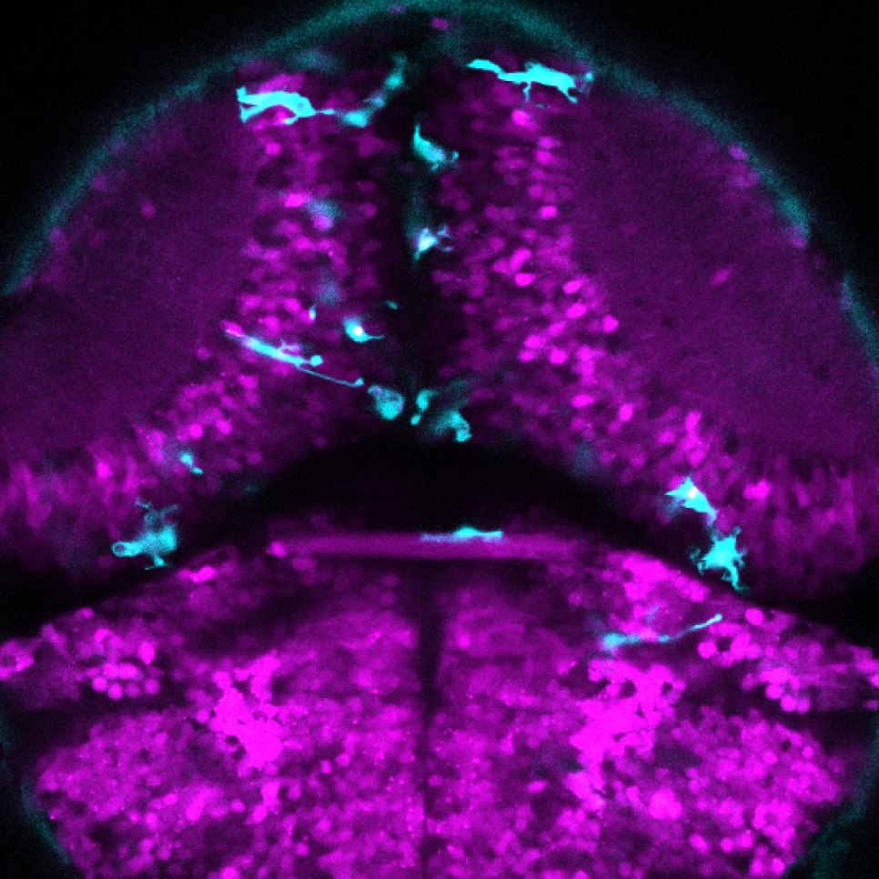
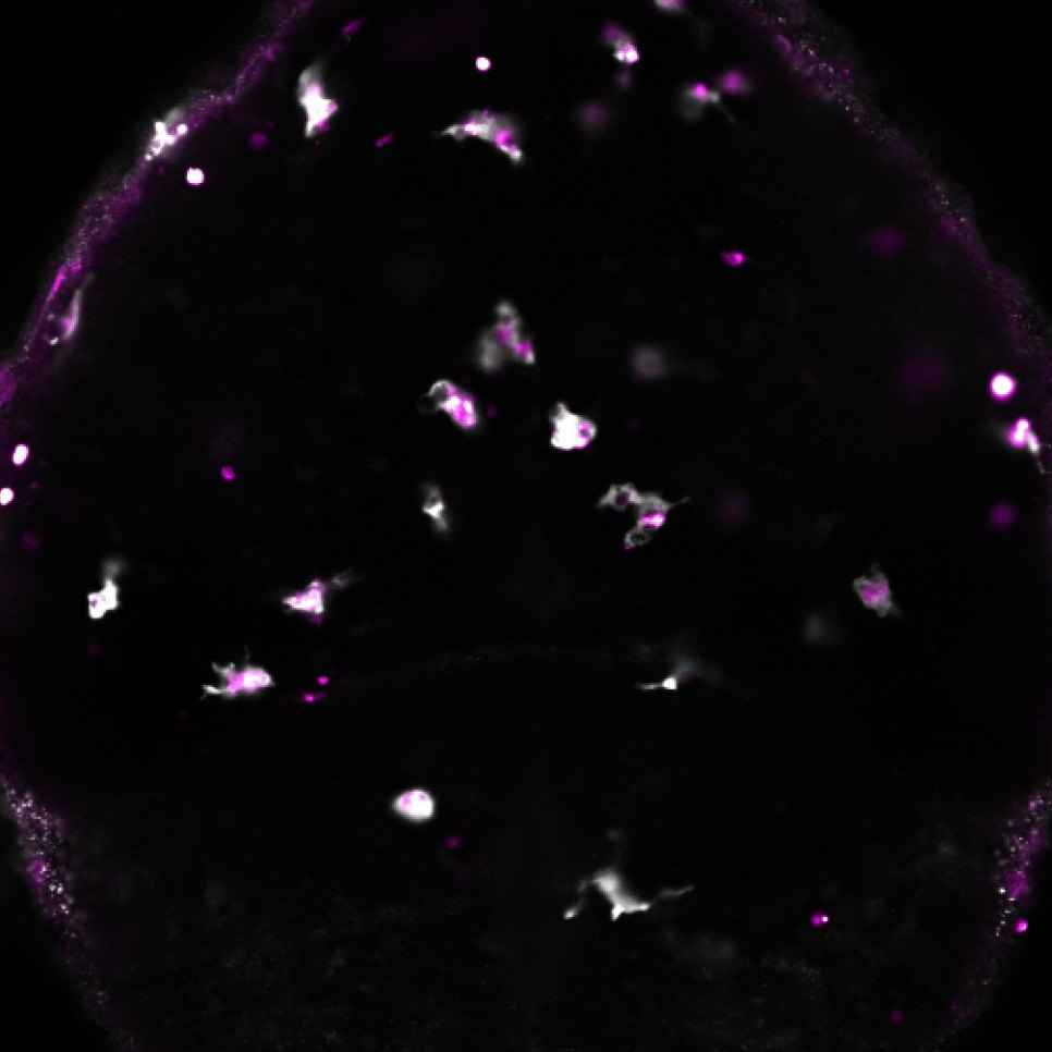
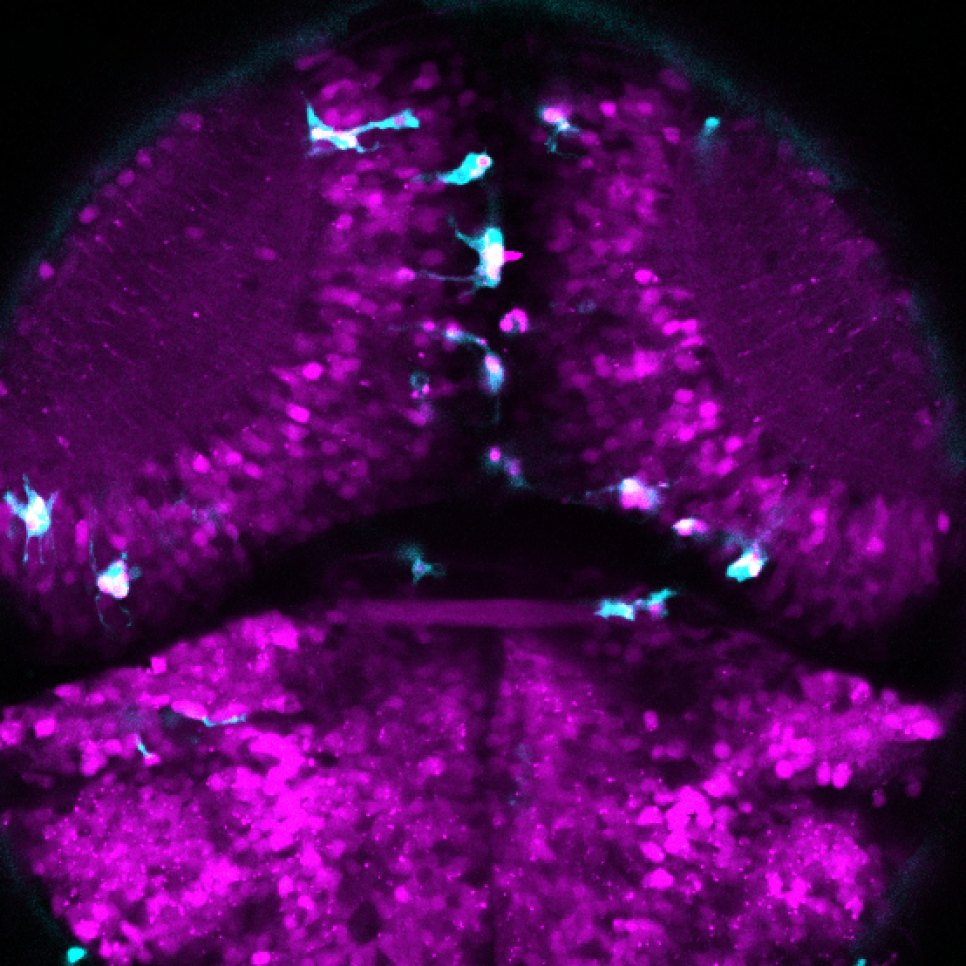
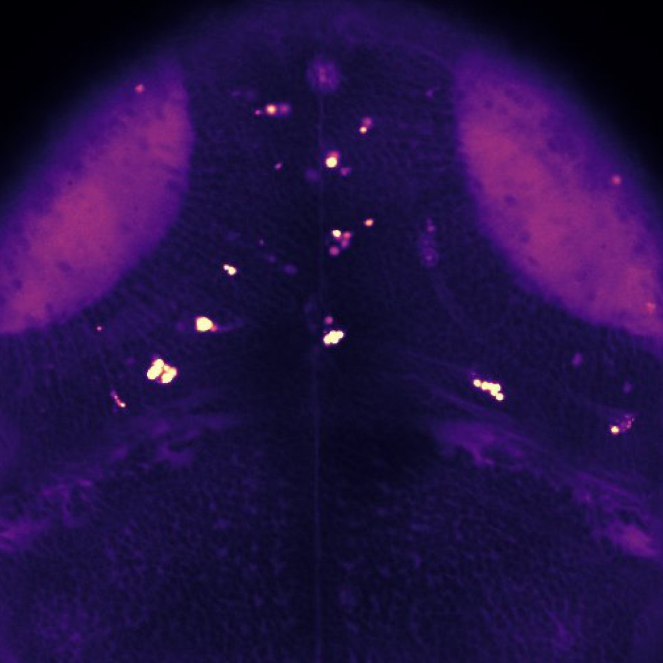
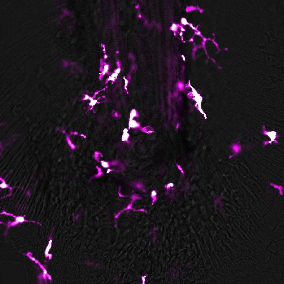
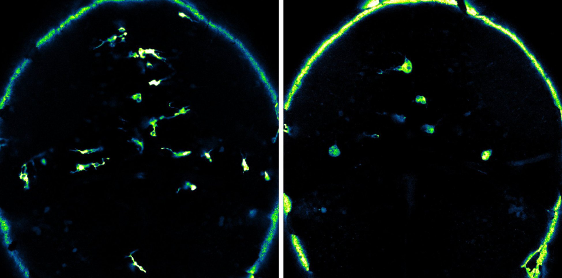
Time-lapse imaging of the zebrafish larval brain, with neurons in magenta and microglia in cyan. In wild-type larvae (left), microglia rapidly survey their environment for any perturbation. In larvae carrying a mutation in an Alzheimer’s disease–associated gene (right), microglia are distended and abnormally rounded; they move little, remaining stationary while extending processes. Using patient-associated variants, we aim to reveal how this mutation, and the resulting disruption of microglial function, contributes to disease pathology. Movie: Emilio Méndez Scolari
Evolution

Evolution of microglia and macrophages.

The Mexican tetra, Astyanax mexicanus, is a natural evolution experiment. Alongside its river-dwelling surface form, A. mexicanus has more than 30 cave populations that adapted to perpetual darkness, sensory deprivation, and food scarcity. Notably, many of the traits that set cavefish apart converge on microglia and macrophages.

In the nervous system, cavefish show eye degeneration, expanded non-visual senses, and rewired neuronal circuits under increased metabolic stress: changes that microglia, which prune synapses and remodel neuronal circuits, are poised to mediate. In the body, cavefish invest less in innate immunity, store more fat, resist starvation, and regenerate poorly. Macrophages act as first responders, scavengers, and tissue remodelers in each of these processes, and even eye degeneration depends on macrophages clearing and remodeling tissue.

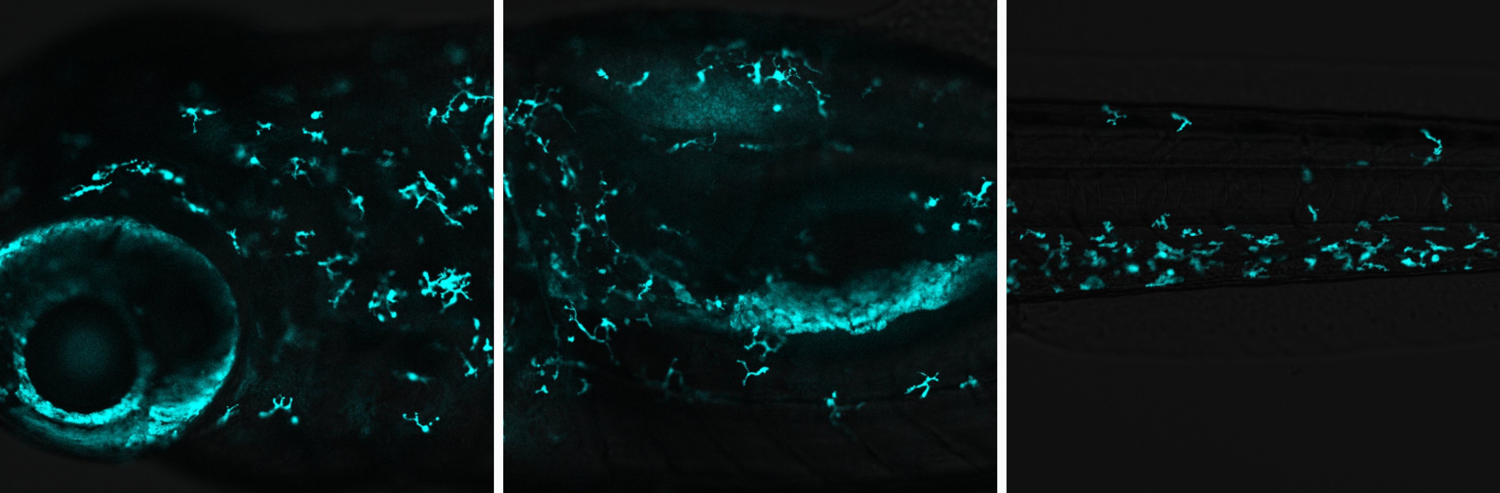
Building on the existing zebrafish genetic toolkit, we developed live-imaging tools for A. mexicanus that have already revealed evolved differences in microglial and macrophage cell biology between surface fish and cavefish. We now ask how microglia and macrophages develop, function, and respond to challenge in cave and surface populations, and what the solutions evolution has found can teach us about these cells in human health and disease.

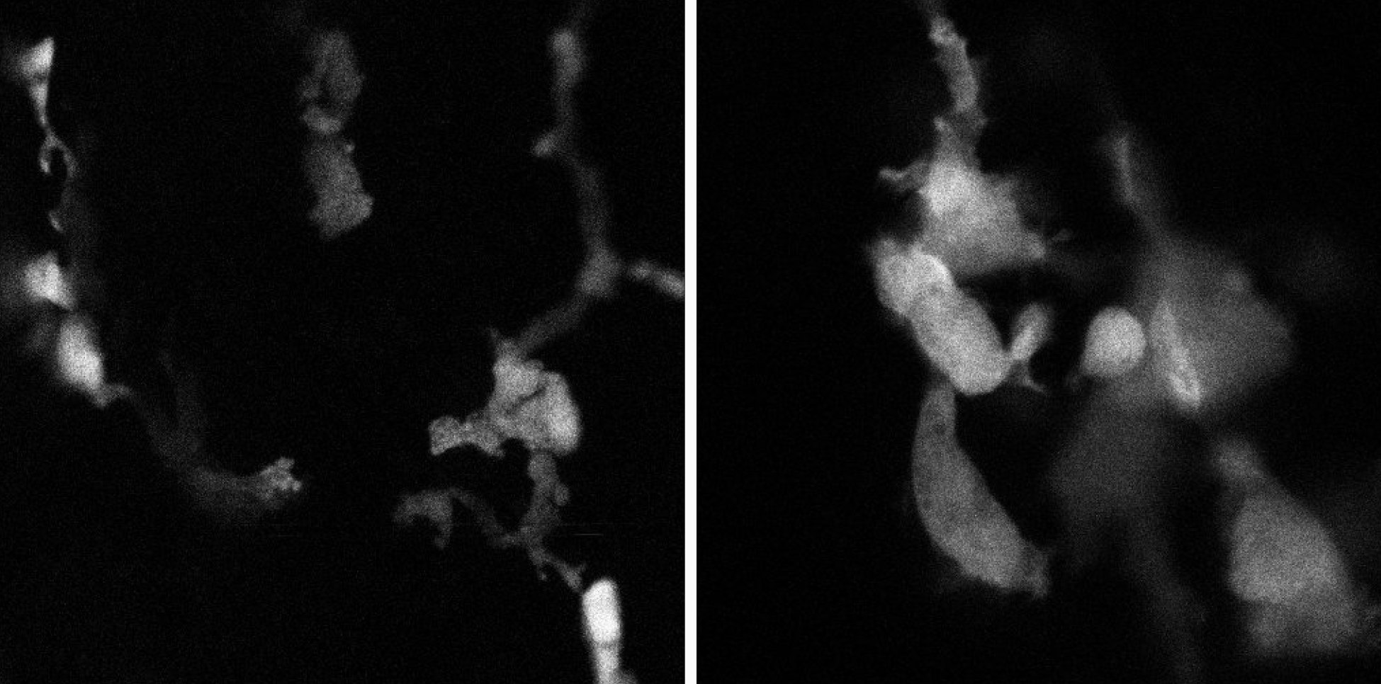
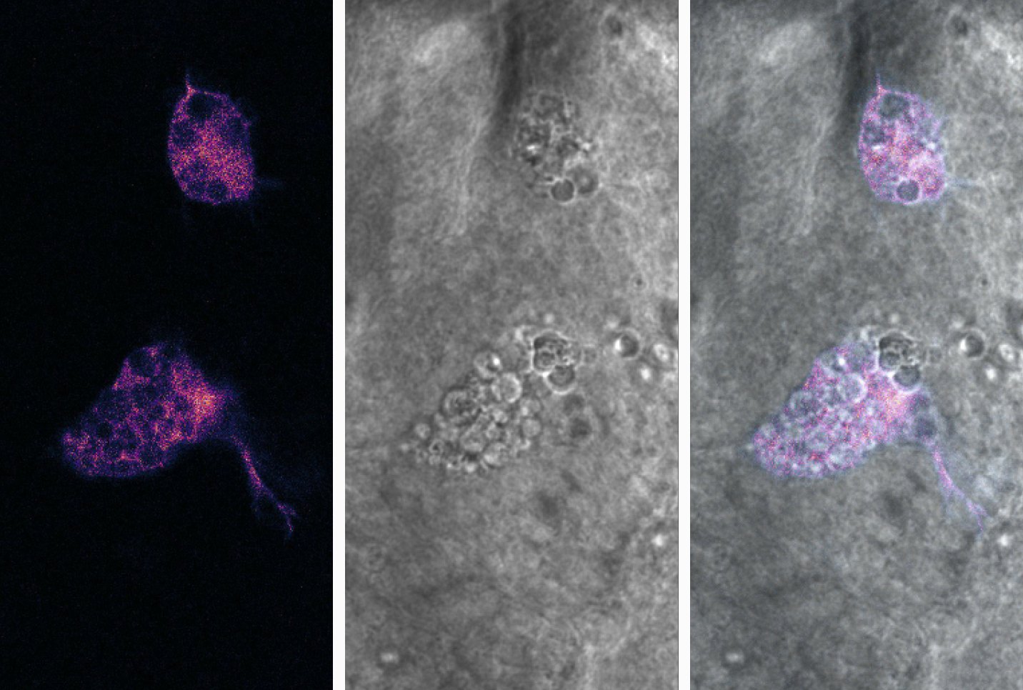
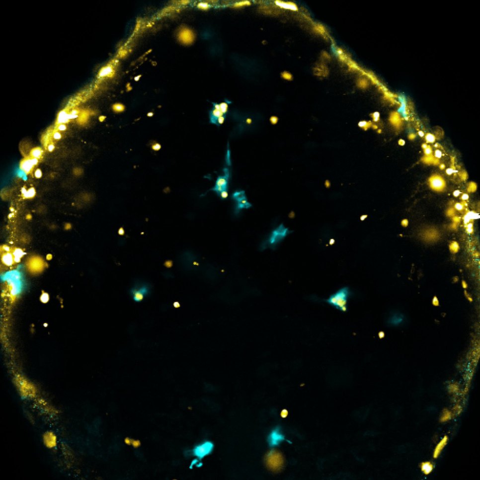
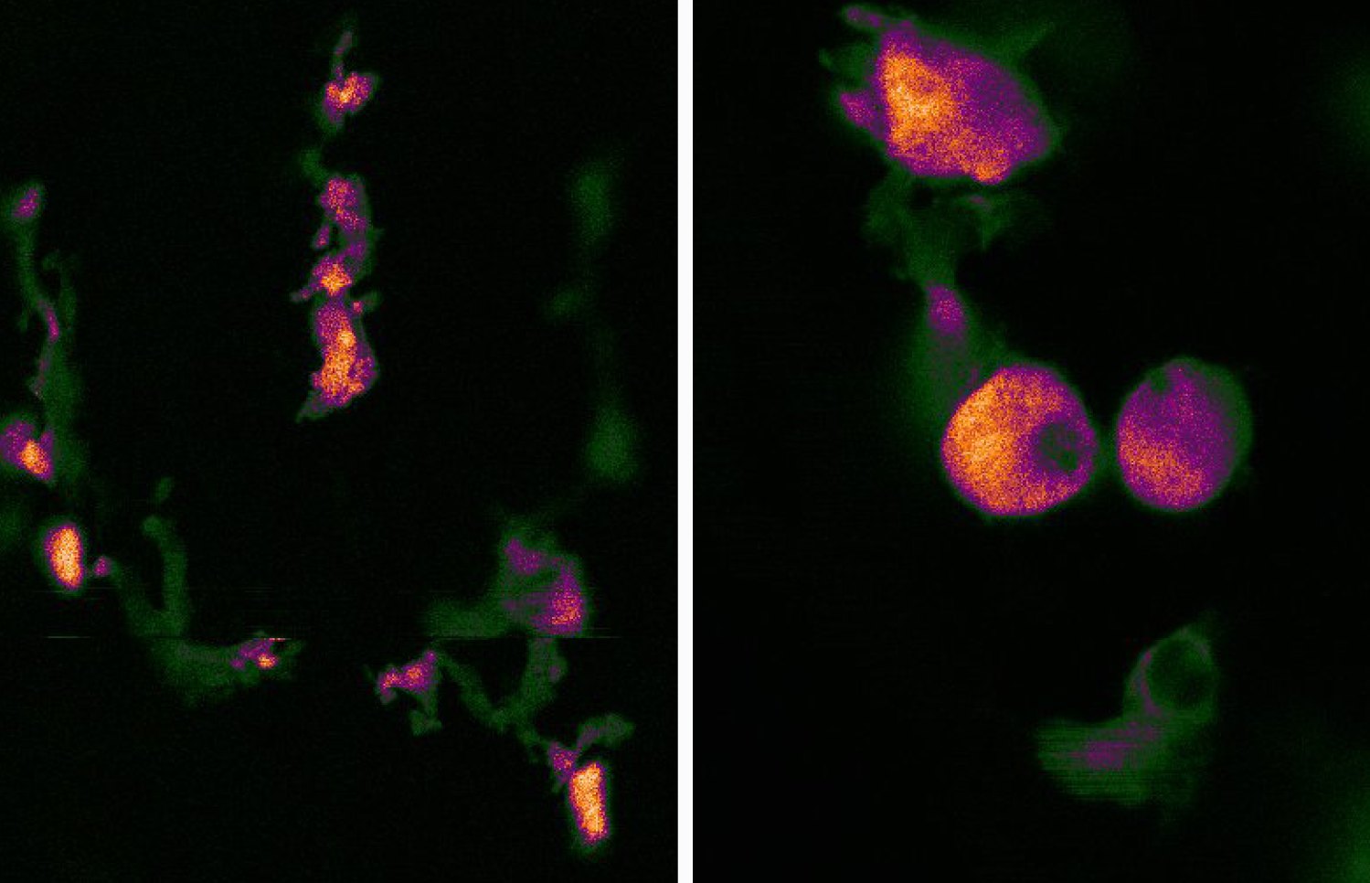
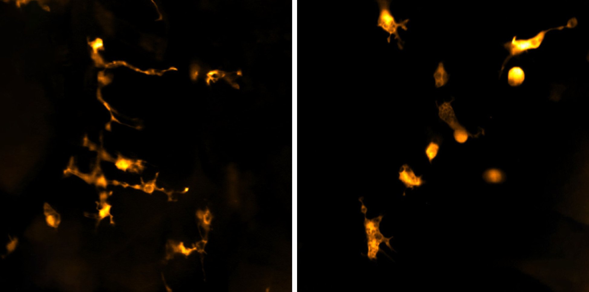
Time-lapse confocal movie showing amyloid beta (grey), injected into the brain parenchyma of a cavefish larva, being rapidly engulfed by microglia and colocalizing with lysosomes labeled with LysoTracker Red (orange). Compared with surface fish, cavefish have more microglia over development, and their microglial lysosomes are more acidic and more proteolytically active. Movie: Emilio Méndez Scolari
Cell biology

Cell biology of microglia and macrophages.

Microglia and macrophages spend their lives clearing debris and responding to danger, tasks that depend on their organelles. Lysosomes break down what the cells engulf, while the ER, Golgi, mitochondria, and peroxisomes govern what they secrete, how they use energy, and how they respond to threats. We study how these organelles are tuned during development and remodeled when the cells face stress.

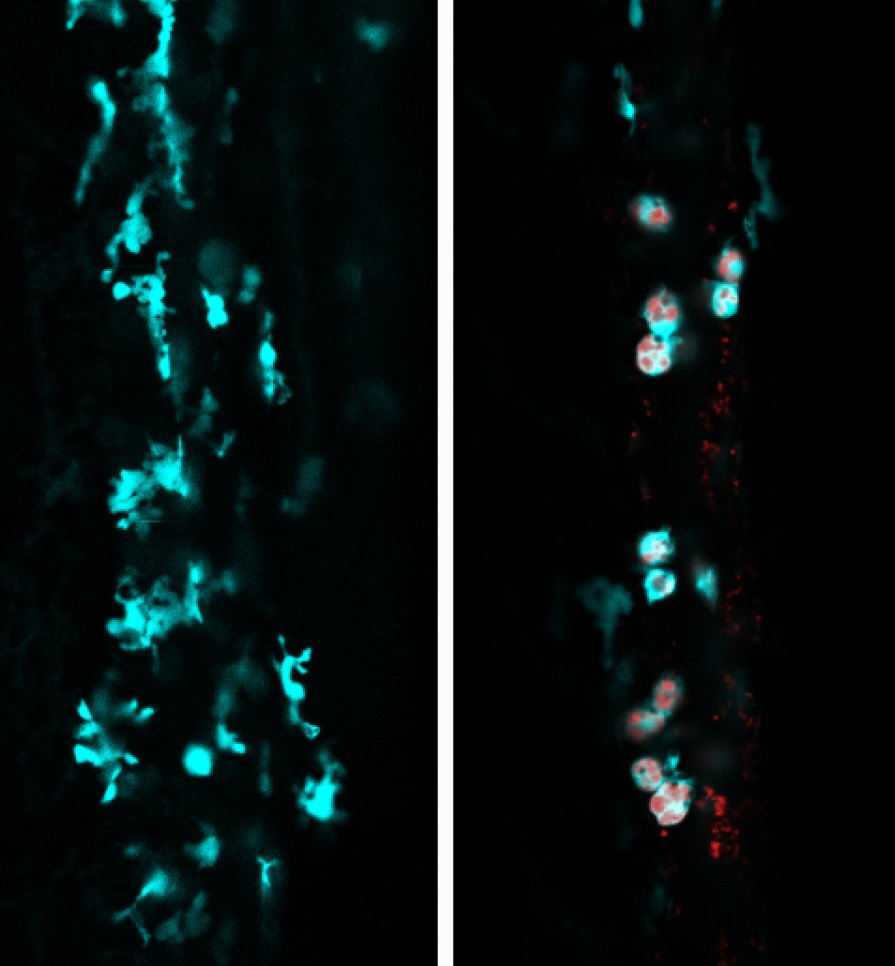
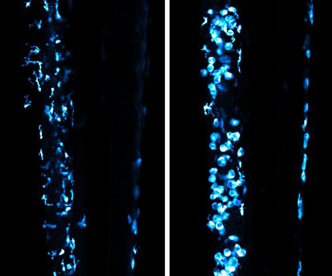
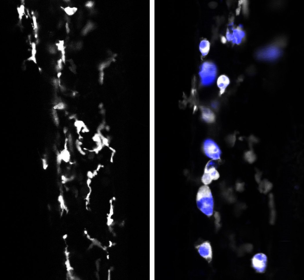
Our earlier work uncovered a lysosomal regulatory circuit that is essential for microglia. The lysosomal regulators RagA and Folliculin keep the transcription factors Tfeb and Tfe3 in check; without them, lysosomal pathways run unrestrained, and embryonic macrophages fail to colonize the brain and become microglia. Surprisingly, Tfeb and Tfe3 are not needed for lysosomes to work under normal conditions, but they are required to switch on lysosomal genes when macrophages are challenged with stress.

These findings raise the central question of this research direction: how are signals of extracellular stress relayed to lysosomes and other organelles? We are working out how microglia and macrophages sense a wide range of environmental cues and transmit them to their organelles, with implications for infection, repair, aging, and neurodegeneration.

Movie
Physiology & behavior

Impact of microglia on animal physiology and behavior.

Microglia act locally, but their effects impact the whole animal. During early development, microglia help build the brain: they eliminate weak synapses, support the birth of new neurons, and clear the many cells that die as circuits form. Because they also sense changes in the environment, microglia are well placed to translate early experiences into lasting changes in brain wiring and behavior.

Early life stress is a major risk factor for psychiatric disease, metabolic dysfunction, and disrupted sleep, and its effects can last long after the stress has ended. With our collaborators, we use a zebrafish model of early life stress to ask how microglia respond during a critical developmental window. We measure how stress and sleep loss alter microglial number, morphology, surveillance, clearance of neuronal debris, and lysosomal function, both during development and into adulthood.

We also ask the reverse question: which circuits depend on microglia in the first place? By transiently disrupting microglia during early development and then letting them return, we can identify the lasting consequences of their absence. Behavioral assays, including tests of stress responses, learning from aversive cues, and sleep, then reveal which circuits and behaviors microglia control, and CRISPR screens point to the microglial genes responsible.

Sensory enrichmentNegative reinforcement
A two-chamber learning maze for zebrafish, with a sensory enrichment zone (red) and a negative reinforcement zone (green) joined by a narrow hallway. Trained fish (bottom) learn to move toward the enrichment zone and avoid the negative reinforcement zone, whereas untrained fish (top) do not. Movies play at 6× real time. Movie: Emilio Méndez Scolari and Brandon Martz